Cardiopulmonary Resuscitation and Emergent Cardiac Care(part ⅠⅠ)
Cardiopulmonary
Resuscitation and Emergent Cardiac Care(part ⅠⅠ)
Prof. Dr. M. Sherif Mokhtar, MD
Prof.of Cardiology,
Professor of Critical Care Medicine
Cairo University
Devices:
Ⅰ.Esophageal-Tracheal Combitube, Laryngeal Mask Airway, and Face Mask Ventilation:
Due to the practical issues of adequate training for endotracheal intubation, the Combitube has been recognized as a reasonable or alternative when endotracheal intubation is not possible or feasible. This device isolates the esophagus and allows for airway isolation for ventilation and avoidance of gastric inflation. One of the most important issues for prevention of potentially fatal complications is the correct identification of the distal port[4].
Laryngeal Mask Airway
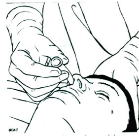
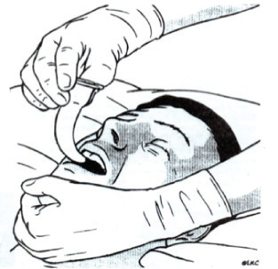
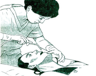
has been shown in many clinical noncardiac arrest scenarios that it is simple to use and provides a safe alternative to intubation. The new guidelines recommend its use in a manner similar to the Combitube, when endotracheal intubation is not possible or feasible. The only disadvantage is that a small percentage of patients cannot be adequately ventilated and an alternative airway management technique should be available. Both of those airway adjuncts have been recommended as a Class IIa intervention in the AHA 2005 guidelines. (fig2)
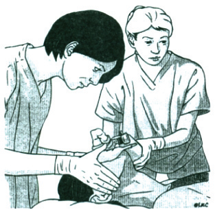
The two-person technique for bag-mask ventilation Laryngeal mask airway
When using a Face Mask, for ventilation, a two-person technique is recommended. One person should maintain the correct head position, the complete seal, and a jaw thrust to maintain airway patency, and the second person should squeeze the resuscitator bag. This approach can be used for a more prolonged period of time when adequate personnel are available, as it enables rescuers to perform high-quality CPR without stopping compressions and interrupting circulation to place an advanced airway device.
ⅠⅠ. Inspiratory Impedance Threshold Device:
The dynamic energy of the expanding chest wall during the decompression phase can be harnessed in order to increase venous return, lower intracranial pressure, and increase circu¬lation to the heart and brain. The inspiratory impedance threshold device (ITD) regulates the entry of air through the airways into the chest during the decompression phase of CPR. It causes a decrease in intrathoracic pressure to -5 to -10 mmHg, and thereby helps to generate a greater intrathoracic vacuum to draw blood back to the heart during the recoil phase of CPR[1].
Although this device is attached to an airway, it is used during CPR to enhance circulation. The ITD has been shown repeatedly to improve blood flow to vital organs and survival in animal and human studies[2]. In clinical studies, the ITD doubles the systolic blood pressure during CPR and increases the chances for short-term survival[3].
Ⅲ. Compression Devices:
The Load Distributing Band (LDB) device (Autopulse, Zoll Circulation), an automated band-compression device, has shown significant improvement in perfusion and pressures in animals and humans[5]. It is based on the physiological principal that circumferential thoracic compression increases intratho¬racic pressure without significant cardiac
produce forward flow.
This device was given a Class IIb level of recommendation, suggesting that it is probably of benefit. A recent randomized trial (ASPIRE) was prematurely stopped because survival to hospital discharge was found to be lower in the LDB-CPR group (5.8% vs. 9.9%) [P = 0.04]. A second study of LDB-CPR, published at the same time as hhe ASPIRE study, however, showed the opposite results.
Compared with resuscitation using manual CPR, a resus¬citation strategy using LDB-CPR on EMS ambulances was associated with improved survival to hospital discharge in adults with out-of-hospital nontraumatic cardiac arrest[6].
Active compression decompression (ACD) CPR devices were recommended with a Class IIb for in-hospital use and Class indeterminate (more data needed) for prehospital use. A small number of in-hospital studies with this device have shown an increase in short-term survival rates[7].
Therapeutic Hypothermia
In two large randomized human trials, mild to moderate hypothermia (32-34°C), post-resuscitation, resulted in an improvement (16-23% absolute risk reduction) for poor neurological outcomes in patients who had a witnessed VF arrest. There was a significant improvement in 6-month survival rates in the hypothermic groups [8,9]. In resuscitated victims of cardiac arrest, especially after prolonged resuscitation efforts, hypothermia should be considered and implemented when possible. The current guidelines have given hypothermia a Class IIa level of recommendation for comatose patients with VF arrest.
For all other presenting rhythms of cardiac arrest, hypothermia was given a Class IIb recommendation. Recent studies have shown that it is possible to cool during CPR before reperfusion is achieved to further minimize tissue damage before it occurs. Cooling during CPR in animals with veno-venous access systems or with cold IV saline and use of ACD CPR plus the ITD may eventually offer a means to rapidly decrease cerebral temperatures during CPR and improve neurological outcomes [10,11].
Based on data in support of therapeutic hypothermia, the guidelines recommend cooling of comatose patients after successful resuscitation when possible, as long as there is a protocol in place to assure careful monitoring of core temperatures and hemodynamics, prevention of shivering, and maintenance of adequate perfusion pressures during the recom¬mended 24-hour period of cooling
Based on data in support of therapeutic hypothermia, the guidelines recommend cooling of comatose patients after successful resuscitation when possible
Vasoactive Medications:
Evidence for the broad use of vasoactive medications during CPR comes primarily from animal studies. There are no placebo-controlled trials that have demonstrated long-term benefit of either epinephrine or vasopressin. As such, the new guidelines recommend the use of either of these agents, with a Class IIb level of recommendation.
Epinephrine is the most commonly used vasopressor during CPR. The beneficial hemodynamic effects of epinephrine during CPR are due to its potent alpha-adrenergic effects. The significant increase in central aortic pressures results in significant increase in coronary and cerebral perfusion pressures and possibly rates of successful resuscitation [12].
However, based on multiple clinical trials, use of high-dose epinephrine is contraindicated and harmful in patients in cardiac arrest (Class III recommendation). The guidelines continue to recommend 1 mg of epinephrine every 3-5 minutes (recommendation Class IIb) for adults in cardiac arrest. If no venous access has been obtained, endotracheal or intraosseous administration can also be effective.
Vasopressin is recommended as an alternative vasopressor during CPR. It too has potent vasoconstricting properties. No study has shown that vasopressin use will result in an increase in hospital discharge rates when used in patients in cardiac arrest. A recent study showed that the Combination of Epinephrine Plus Vasopressin resulted in higher rates of return of sponta¬neous circulation, no increase in long-term survival rates, but a strong trend toward worsening of neurological outcomes, except in those with an initial rhythm of asystole arrest [13].
There is no good treatment for asystole. Atropine, a vagolytic medication, has no known untoward effects in patients with asystole, and can be given for severe bradycardia and asystole, with doses of 1 mg i.v. every minute to a total dose of 3 mg (Class Indeterminate). There is no randomized animal or human
study to support the administration of atropine for improvement of outcomes.
Antiarrhythmic agents:
As with the other IV medications, there were insufficient levels of data or consensus among the experts regarding the use of antiarrhythmic agents during CPR.
Amiodarone is now considered the drug of choice and as an IV bolus of 150-300 mg for VF or pulseless VTs that are unresponsive to the initial sequences of CPR-shock- CPR-vasoconstrictors. The recom¬mendation is based on limited clinical trials [14,15] showing improvement in hospital admission, but no definitive increase to hospital discharge rates, when compared to placebo or lidocaine.
Given the lack of definitive data, Lidocaine (initial dose of 1-1.5 mg/kg IV) can also be used in patients in cardiac arrest (Class Indeterminate)
Reperfusion therapy
Due to the high incidence of obstructive CAD (70% of autopsy patients document active thrombus in the coronary tree and from patients undergoing cardiac catheterization, another 70% show evidence of severe coronary artery stenosis) in the cardiac arrest population and the inability to make the diagnosis of STEMI, based on the post-resuscitation ECG, elective angiography and primary angioplasty should be considered in all survivors without any other clear etiology for the cardiac arrest [16].
- Application of good-quality CPR, limitation of compression interruptions and ventilations, and addition of an ITD, early post-resuscitation hypothermia and reperfusion therapies have shown promise when implemented as a system rather than unique interventions [17].
- 2. Adjuncts to improve circulation during CPR, such as the ITD, have also been recommended as higher than vasoactive medications.
- 3. Comatose patients with VF arrest could be treated with mild therapeutic hypothermia as long as the infrastructure needed to support the patients is promptly available.


 info@utopiapharma.com
 Plot No. (2) Industrial Zone (A7) - formerly Zizinia - Cairo - Ismailia Road - 10th of Ramadan - Sharkia